Recipe for organ tissue creation
Posted by magazine
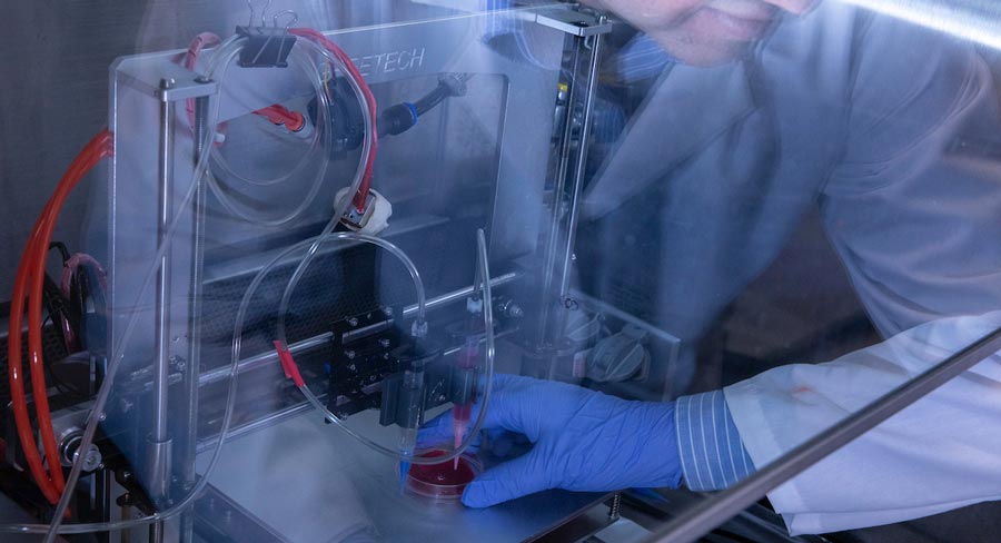
Bioactive glass is added to the hydrogel/cell mixture and loaded as “bio-ink” into a 3-D printer, where it then prints the cells onto a lattice in a petrie dish.
Using bioactive glass, stem cells and a 3-D printer, Missouri S&T researchers are creating organ tissue samples in hopes of advancing pharmaceutical testing and providing a better understanding of how diseases affect human cells.
The researchers grow stem cells and add them to hydrogels made of alginate, gelatin or similar substances. Then, in a step unique to Missouri S&T, the researchers add bioactive glass to supply needed calcium ions to the hydrogel/cell mixture and load the mixture as “bio-ink” into a 3-D printer. They test the samples after fabrication to assess the stem cell function, the material’s tensile strength, degradation and the best glass type to add.
“Different cells prefer different gels, so we work to find which gel combination suits our research.”
“Different cells prefer different gels, so we work to find which gel combination suits our research,” says Krishna Kolan, MS ME’11, PhD ME’15, a postdoctoral researcher in mechanical and aerospace engineering. “The challenge is that dissolved glass adds calcium, but it changes the pH, and cells need neutral pH to survive. We figured out which glass and how much to add to maintain neutral pH.”
Kolan says researchers are several years away from making a functioning organ, such as a liver or kidney. The challenge is the vascular system and multiple types of cells in those organs, but S&T researchers are working on ways to develop vascular systems within the bioprinted tissue. Undergraduate students August Bindbeutel in mechanical engineering and Lesa Steen in materials science and engineering are assisting Kolan.
“Endothelial cells form networks in environments they like, such as glass-infused hydrogel,” Kolan says. “As the network grows, it vascularizes the tissue.”
As researchers work toward someday repairing or replacing organs with engineered organs, they create tissue models for pharmaceutical testing, Kolan says. His team is also working on 3-D-printed bone models. Biology graduate student Bradley Bromet is comparing diseased cells with healthy stem cells to see in 3-D how a disease — diabetes, for instance — affects cells.
Kolan is working on the project with Ming Leu, the Keith and Pat Bailey Professor in S&T’s mechanical and aerospace engineering department; Richard Brow, interim deputy provost and Curators’ Distinguished Professor of materials science and engineering; Delbert Day, CerE’58, Curators’ Distinguished Professor emeritus of ceramic engineering, and Julie Semon, assistant professor of biology and director of S&T’s Laboratory of Regenerative Medicine.